Image number: 36a
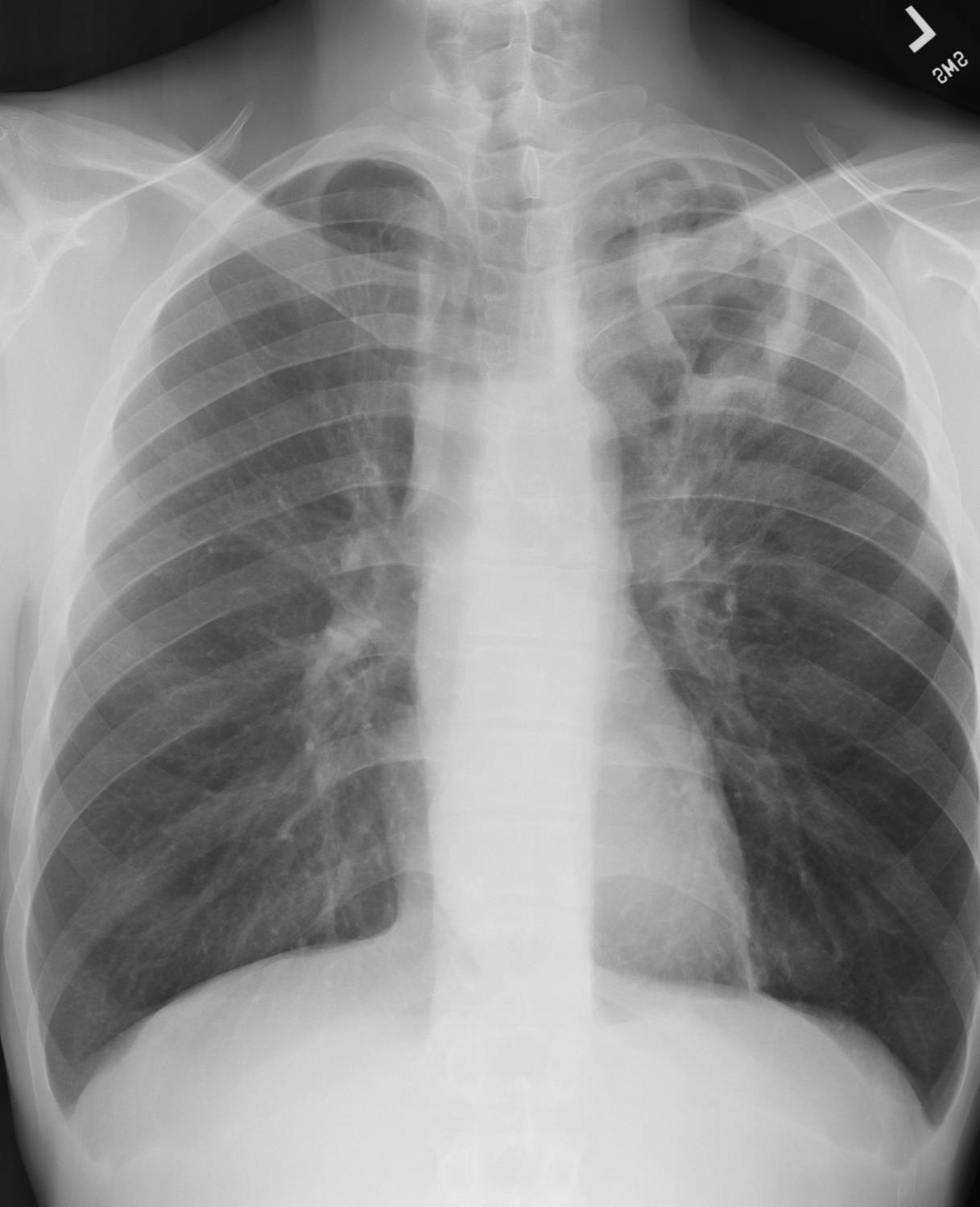
Image number: 36b
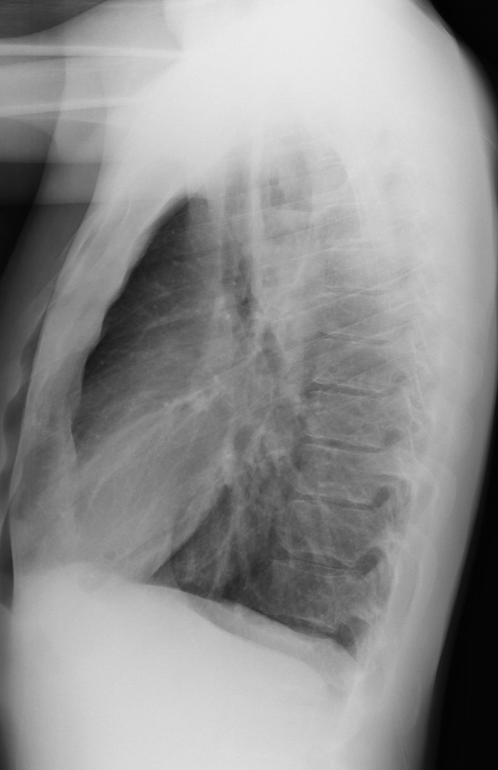
Image number: 36c
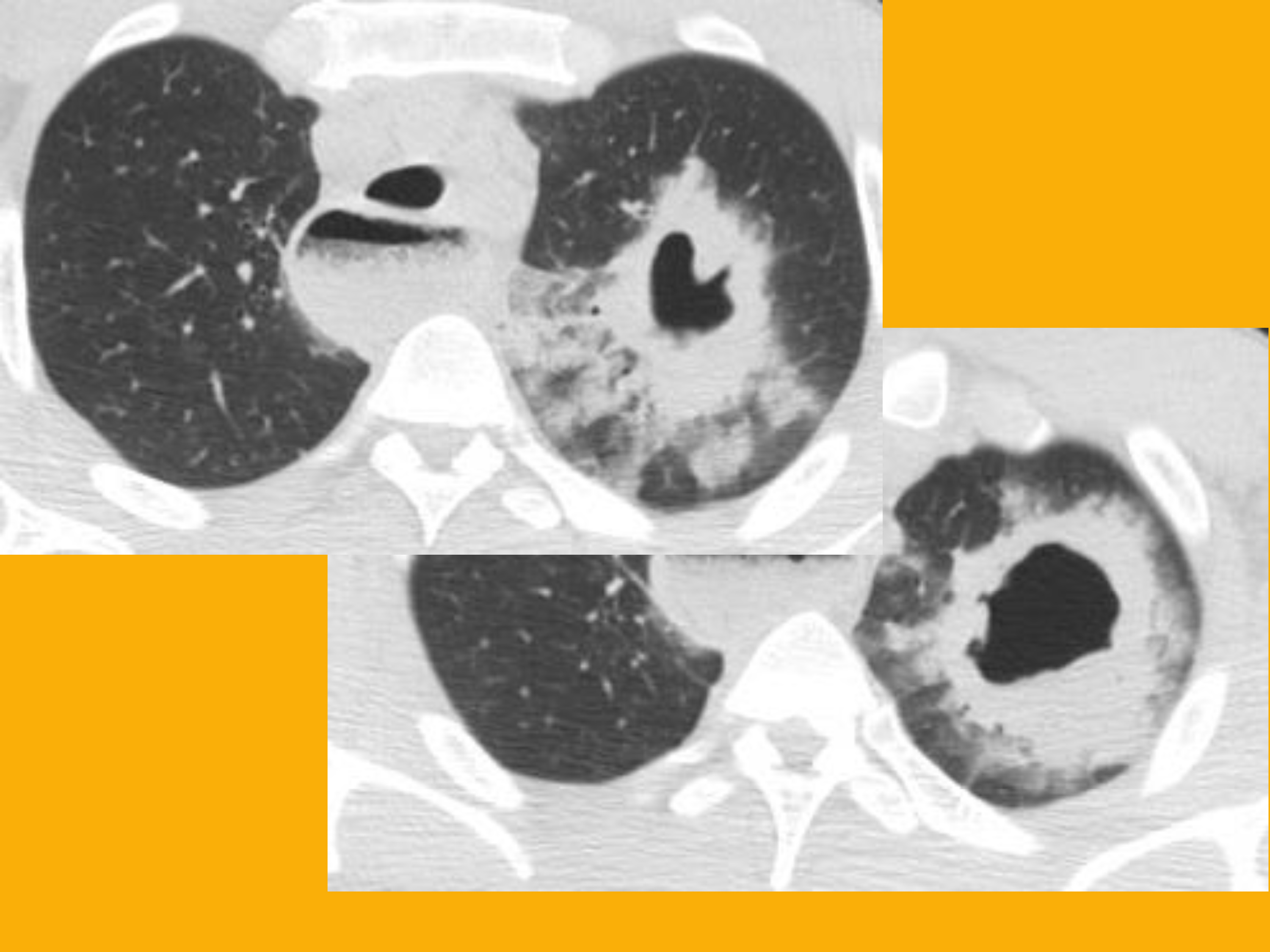
Image number: 36d
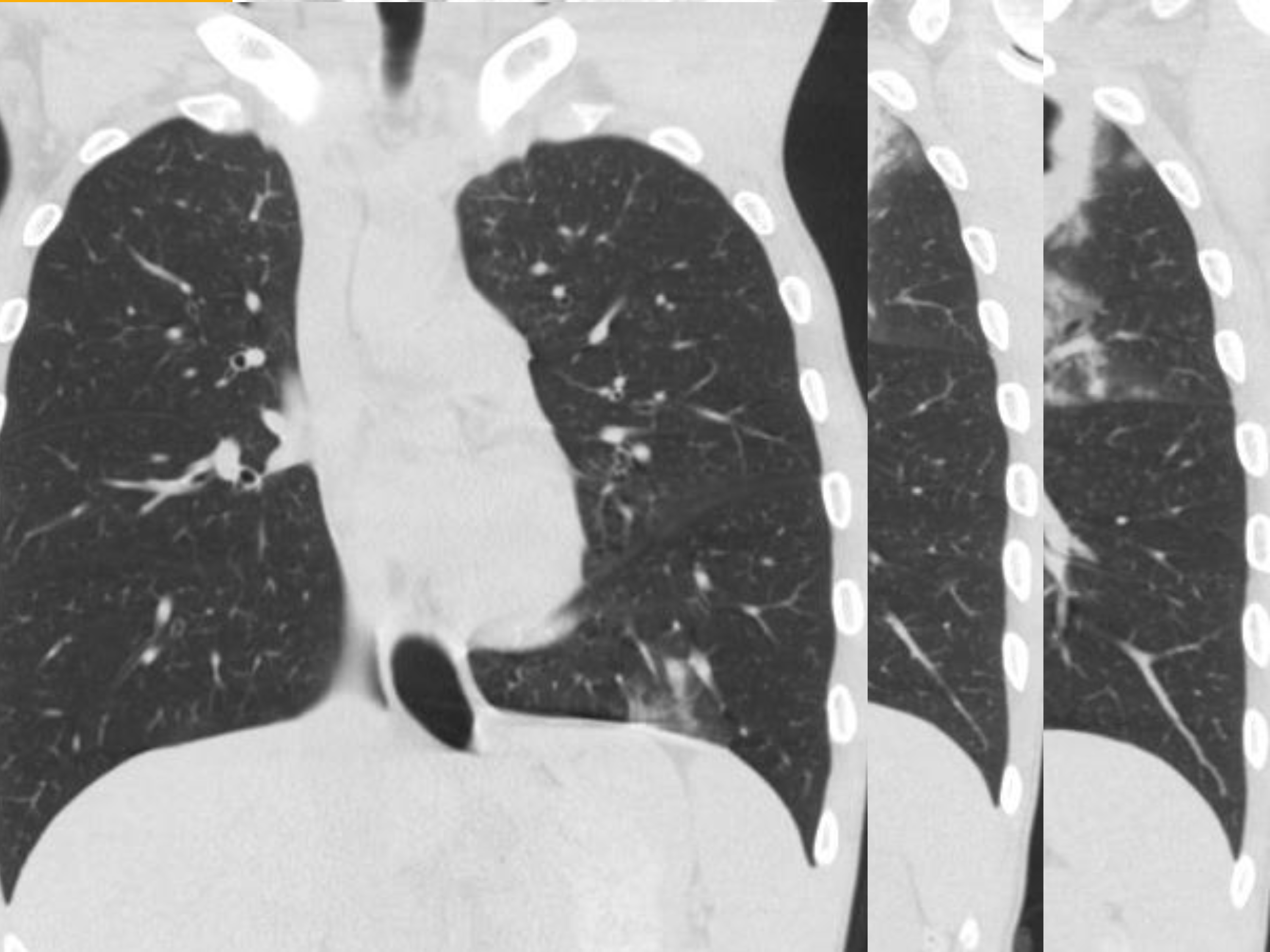
Image number: 36e
Category:
Other non-TB
Description:
36a - Large left apicoposterior irregularly border cavity containing a small air-fluid level.
36b - Large left apicoposterior irregularly border cavity containing a small air-fluid level.
36c - Axial CT images show a large thick-walled cavity with surrounding ground-glass and consolidation.
36d - Coronal CT images show a large thick-walled cavity with surrounding ground-glass and consolidation.
36c & 36d - A markedly dilated esophagus filled with air-debris level consistent with achalasia is noted. The cavity is an anaerobic abscess likely from aspiration.
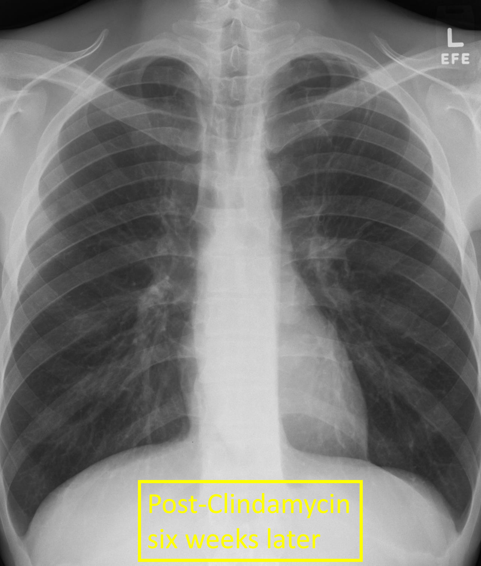
36e - Interval decrease size and wall thickness of left upper lobe cavity after six weeks of Clindamycin.
Content Provider:
Thienkhai Vu, MD