TB Radiology Resource Page
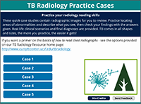
These brief, interactive self-study case studies allow clinicians to practice locating areas of radiographic abnormalities using real-life clinical scenarios. This product was produced in collaboration with the Firland Northwest Tuberculosis Center. 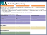
An open-access resource library of radiographic images of tuberculosis. Individuals may use these images to gain an appreciation for the broad spectrum of radiographic manifestations of tuberculosis. The images are free to download and share for non-commercial educational purposes. This product was produced in collaboration with the Firland Northwest Tuberculosis Center. 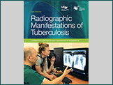
Using a self-study format, this publication presents the basic principles of how radiographs capture images, normal chest radiographic anatomy and interpretation of disease patterns. The radiographic presentations for tuberculosis are covered in detail and reinforced with clinical case exercises. A print version of this textbook can be ordered or an electronic version downloaded online. 
A 54-minute self-study module, this online tool allows learners to listen as CITC's medical director, Lisa Chen MD, presents the basics of how to read chest radiographs: covering basic anatomy, disease patterns, and tuberculosis specific presentations. Learners can listen or read along at their own pace.  
The animated teaching slide-set (adapted and used for the audio-visual presentation above) is available here in a version first developed as part of a set of teaching modules for the International Standards for Tuberculosis Care (ISTC). The teaching notes are more detailed than in the self-study version to help prepare the presenter with background information for training as well as offering interactive teaching suggestions and slide-show animation cues. Basic Chest Radiology for the TB Clinician | | Date: June 18, 2018 
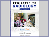
This 60-minute webinar presentation (followed by a 15-minute Q & A session) is the continuation of the Pediatric TB Radiology webinar Part 1 (May 2017) which focused on the diagnosis and follow-up of pediatric cases along with reading of the chest x-rays. Date: May 5, 2017 
This webinar was created for physicians, nurses, and other health professionals who diagnose and treat patients with TB. The training focused on the diagnosis and follow-up of pediatric cases along with reading of the chest x-rays. 
Date: July 16, 2012 Originally presented in July 2012 by Brett Elicker, M.D. and Lisa Chen, M.D., this 90-minute webinar covered the basics of interpreting chest radiology for findings of tuberculosis. |
| |
|
This book for clinicians shows and describes examples of radiographic abnormalities common in pediatric tuberculosis, emphasizing pulmonary, lymphatic and meningeal disease. The utility of CT scan and MRI in pediatric TB are also discussed. Radiographs and case studies are used as illustrations throughout the book. |